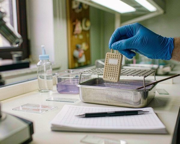

# What Consumables Does Histology and Tissue Staining Need?
Walk into any histopathology lab and the machines get all the attention: the microtome, the tissue processor, the stainer. But the consumables decide whether a 4 µm section survives the day. This guide walks through the histology consumables a working lab actually burns through, and why each one earns its place on the order list.
Histology needs embedding cassettes and molds, microtome blades and forceps, charged microscope slides, staining racks and dishes, reagent and stain bottles, filters, nitrile gloves, and tissue-processing consumables. Every one of them protects section quality, stain consistency, and diagnostic reliability.
If you do the ordering, or if sections keep lifting off your slides, the detail below will save you a re-run.
The Full List, Section by Section
Cassettes and embedding consumables
Everything starts at grossing. Tissue goes into a cassette, small biopsies into perforated biopsy cassettes, larger specimens into standard ones with a foam pad or biopsy paper to stop fragments slipping through. From there the cassette rides through fixation and processing and ends up embedded in paraffin inside a metal mold.
So the shopping list here is longer than people expect: cassettes and lids, embedding molds or rings, processing/embedding paraffin, biopsy pads, and embedding paper. Fixation itself deserves respect. The classic 10% neutral buffered formalin works because formaldehyde crosslinks proteins in a well-understood way, something Fox and colleagues laid out decades ago and labs still rely on. Under-fixed tissue haunts you later as cracks, holes, and stains that refuse to behave.
Microtomy: blades, brushes, forceps
The microtome cuts, but the blade does the work. Most labs use disposable high-profile or low-profile blades and change them far too late. A dull blade gives compression, chatter, and “venetian blind” scores. My rule of thumb: one blade per block for routine work, and never stretch a blade across a calcified specimen.
Around the blade you need fine forceps (hinting forceps are easier on the wrists), a natural-hair brush for guiding the ribbon, blade removal tools so nobody grabs a used blade bare-handed, and an ice bath or cold plate to chill blocks. Warm tissue smears and folds; cold tissue cuts clean. Leica’s guide to specimen preparation covers this workflow well if you want the full picture.
Slides: plain, charged, and everything between
A section floating on a 42 °C water bath looks fragile, because it is. Getting it onto glass without wrinkles is half the job; keeping it there through dewaxing, staining, and antigen retrieval is the other half.
Routine H&E gets along fine with plain or frosted slides. But immunohistochemistry (IHC) and in situ hybridization subject slides to heat-induced epitope retrieval (HIER), often at high pH, and plain glass loses tissue at exactly that moment. Positively charged slides such as Superfrost Plus microscope slides hold sections electrostatically and chemically through the whole barrage. If your lab sends any tissue for IHC or molecular testing, this is not an upgrade. It is the baseline.
I learned that the expensive way. Years ago, a set of lymph node sections lifted clean off the slide mid-run during HIER, every single one. The cause: a tech had grabbed plain slides from an old box at the back of the cupboard. We re-cut the whole case, re-ran the stainer, and lost a full day. Two boxes of charged slides cost less than that afternoon.
Staining: H&E reagents, bottles, and filtration
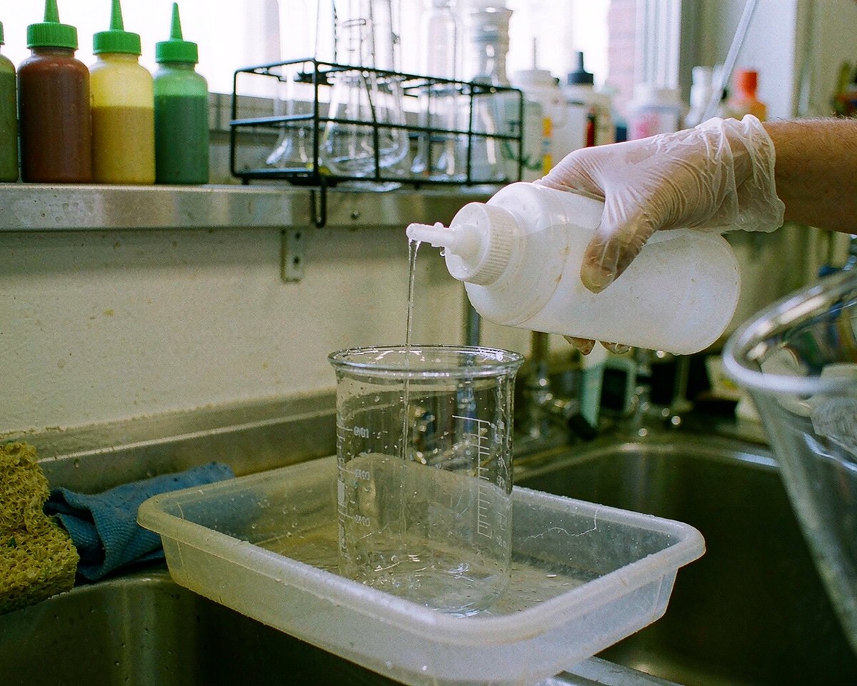
H&E looks simple and is anything but. You are managing hematoxylin (nuclear stain), acid alcohol or differentiator, bluing reagent, eosin (cytoplasmic stain), and graded alcohols with xylene, all in open baths that drift every day. Each bath lives in its own reagent bottle or staining dish, and the bottles need to be pour-friendly and chemical-resistant.
Two consumable habits keep H&E reproducible:
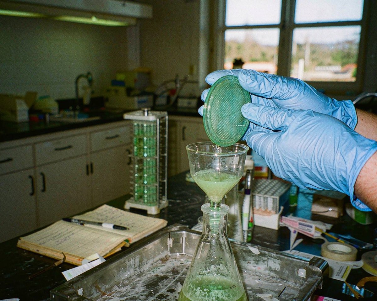
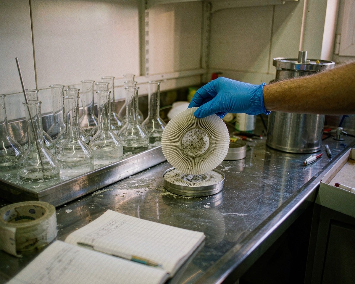
• Filter the hematoxylin. Oxidized hematoxylin and floaters form a metallic scum that deposits on sections. Filter paper and a funnel, once a day or at least a few times a week, depending on throughput.
• Cover baths when idle. Dust, evaporation, and oxidation all slow down with a lid. Reagent bottle caps belong on the bottles too.
Ready-made certified stains like Harris modified hematoxylin solution take some of the mixing variables out of your hands. For special stains such as Giemsa, light is the enemy, and that brings us to storage.
Coverslipping
After staining, a coverslip and mounting medium protect the section for decades. You need coverslips matched to your slide size, mounting medium, and clean xylene for clearing. Skimp here and you get bubbles, hazy media, or a slipper that jams on a folded corner.
Contamination control and gloves
Formalin is a carcinogen and a skin sensitizer; NIOSH’s formaldehyde guidance is blunt about exposure limits. Nitrile gloves, changed often, are the floor. Face protection while handling formalin and xylene, and disposable wipes at grossing, round it out.
One more thing: tissue that moves on to PCR or RNA work picks up everything your consumables carry. If your lab feeds a molecular bench, DNase- and RNase-free consumables stop the trace enzymes that shred nucleic acids. Most routine histology work does not need that grade, so we cover it below.
Choosing Slides for IHC vs Routine H&E
If you standardize on one slide type, make it charged. Plain slides are cheaper per box, but every fallen section costs a re-cut, a re-run, and pathologist patience. Charged slides handle routine H&E without side effects, so the only real cost is a few cents per slide.
For high-pH HIER protocols or particularly uncooperative tissues (bone, brain, breast), ask your slide supplier which of their charged products tolerates your retrieval conditions. Not all charged coatings are identical.
Keeping Your Stains Consistent
Stain baths age with use, and they age differently by bath. Track slides-per-bath counts for hematoxylin and eosin instead of changing everything on the calendar. Filter hematoxylin often, watch for the blue-black scum, and top up rather than replace when a bath is only diluted.
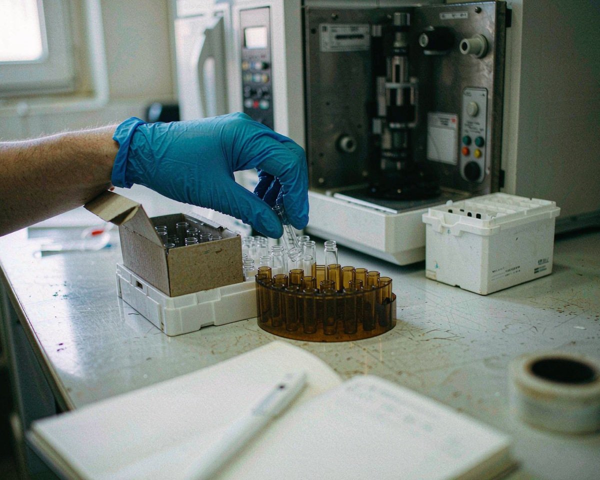
Stain storage matters as much as stain use. Giemsa and other light-sensitive stains fade in clear glass, which is why amber vs clear reagent bottles is a decision worth making deliberately rather than by accident.
Buying Tips for Histology Consumables
A few things I would tell anyone building this order list from scratch:
• Buy blades and slides in one brand per batch where you can. Mixed lots behave differently at the water bath and at the blade holder, and troubleshooting gets murky when four suppliers are in play.
• Check the storage side of the house. Slides and stains have shelf lives, and how you store lab consumables decides whether the last box in the case performs like the first.
• Ignore “sterile” on most histology items. Histology is not cell culture; plain non-sterile slides and cassettes are the norm. The difference between sterile, non-sterile, and low-bioburden claims is worth knowing so you do not pay for a label you do not need.
• Ask for lot numbers and certificates up front. When a stain goes strange, the first question any supervisor asks is what changed.
Conclusion
Histology and tissue staining run on a longer consumable list than any other bench I have worked with: cassettes, molds, paraffin, blades, brushes, forceps, charged slides, stain bottles, filter paper, coverslips, and a steady supply of good gloves. None of these items is glamorous, and every one of them shows up in the final slide. The pattern I would leave you with is simple: consumables that touch the tissue directly (slides, blades, cassettes) deserve your quality budget first, while storage and handling items pay you back in consistency. If you are deciding what to standardize on next, start with amber vs clear reagent bottles for your stain shelf, then review consumable storage practice across the whole department. Small purchasing decisions, repeated weekly, are what keep a histology lab’s slides diagnostic.
Frequently Asked Questions
What are charged slides used for in histology?
Charged (positively charged) slides hold tissue sections in place through harsh steps like heat-induced epitope retrieval in IHC. Plain slides are fine for routine H&E, but charged slides prevent tissue loss during aggressive staining protocols.
How often should hematoxylin be filtered?
Filter it at least weekly in a low-throughput lab, and daily in a busy one. You are removing oxidized dye scum and debris that otherwise deposit on sections as dark specks.
Do histology consumables need to be sterile?
Almost never. Histology fixes and stains tissue rather than culturing it, so non-sterile slides, cassettes, and bottles are standard. Sterile grades matter when material feeds molecular testing or cell culture downstream.
Why are some stain bottles amber instead of clear?
Amber glass blocks the light that bleaches light-sensitive stains such as Giemsa and some eosin formulations. If a stain is stored for weeks between uses, amber storage noticeably extends its working life.
What section thickness is standard for H&E?
Most diagnostic labs cut paraffin sections at 3 to 5 µm, with 4 µm as a common default. Thinner sections show more nuclear detail but tear more easily, so blade condition and water bath temperature matter more as thickness drops.